
Welcome to
Multi CBCT Solution
The only CBCT center run by Dental & Maxillofacial radiologist who are operating this center with proper reporting. They are the only Dental and Maxillofacial radiologist in Bangladesh

Dr. P. C. Mallick

It is our great honor and pleasure to introduce Dr. P. C. Mallick, who is the Chief Consultant of Mallika Dental. Who has passed B.D.S in 1989 from D.D.C under Dhaka University with the place in merit list, got the prestigious Scholarship of ‘Japan Govt. Monbukagakusho’ in 2000 and completed PhD in Hiroshima University, Japan in Dental Science in 2005. Then he joined as a teacher in Pioneer Dental College. After that, he passed B.C.S and started working at Dhaka Dental College and Hospital (DDC&H). At present, he is an Associate Professor in DDC&H.

Dr. Samir Banik
Dr. Samir Banik completed his Bachelor of Dental Surgery from the University of Dhaka, Bangladesh in the year 2001. After that, he joined Bangabandhu Sheikh Mujib Medical University for his post graduation training in the dept. of Prosthodontics. Then he joined Bangladesh Civil Service (BCS) as a dental surgeon. At the same time, he was blessed by a prestigious scholarship from Govt. of Japan (Monubukagakasu). Then he joined Hiroshima University, Japan in the dept of Oral & Maxillofacial Radiology as a Ph.d fellow. He finished Doctor of Philosophy in dental science in the year of 2010. Returning from Japan, he joined Dhaka Dental College and Hospital in the dept of Oral and Maxillofacial Radiology as Assistant Professor. He is a specialist in Maxillofacial Radiology.
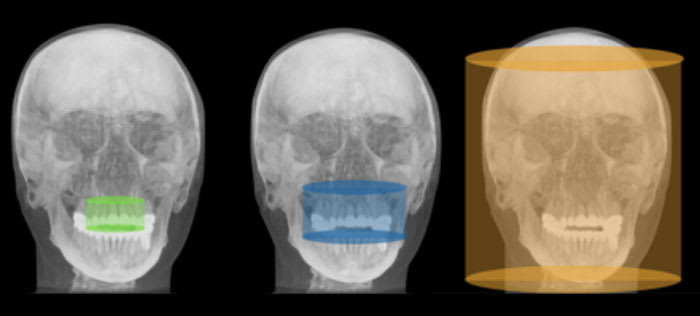
Principles of CBCT
CBCT units can be categorized according to patient positioning, field of view, clinical functionality, and detector type. Clinicians should consider all these characteristics prior to purchasing a unit. The first CBCT unit was similar in design to MDCT, with the patient in a supine (reclined) position during the scan. Most CBCT units have the patient seated or standing; this type of unit requires less space in the dental office and may be more accessible for wheelchair users.1,2 Field of view (FOV) is another way to categorize these units. FOV refers to the anatomical area that will be included in the data volume, or the area of the patient that will be irradiated.7,9 Depending upon the type of machine/detector, and the geometry of the x-ray beam, the FOV could be classified as small or limited, medium and large (Figure 1).